Nervous System Diagram Labeled : File:Nervous system diagram-bn.svg - Wikimedia Commons : Essentially, nerve cells, also known as a neurons, are the active component of the nervous system.
Nervous System Diagram Labeled : File:Nervous system diagram-bn.svg - Wikimedia Commons : Essentially, nerve cells, also known as a neurons, are the active component of the nervous system.. Illustration of peripheral nervous system, medical vector illustration diagram with brain, spinal cord and full body nerve scheme. Understand faster with this handy nervous system revision guide full of quizzes and labeled diagrams. They receive data and feedback from the sensory organs and from. The nervous system is made up of vast neural networks; Be sure to visit the guide for more context and information about full nervous system diagram labeled, or read some of our other health & anatomy posts!
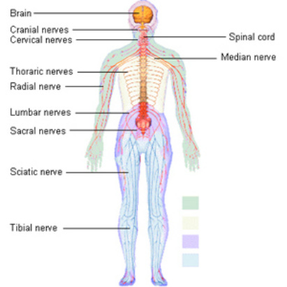
From wikimedia commons, the free media repository. Together, the central nervous system (cns) and the peripheral nervous systems (pns) transmit and process sensory information and coordinate bodily functions. Illustration of peripheral nervous system, medical vector illustration diagram with brain, spinal cord and full body nerve scheme. Please note the obturator nerve has been incorrectly labeled. The central nervous system (cns) and the peripheral nervous system (pns).
File:Nervous system diagram-hi.svg - Wikimedia Commons from upload.wikimedia.org Please note the obturator nerve has been incorrectly labeled. This system consists of all the neuron cell bodies and processes located outside the brain and spinal cord. Learn vocabulary, terms and more with flashcards, games and other study tools. Structure of a typical neuron. Neurons or nerve cells are the basic building blocks or units of the nervous system. Human eye anatomy, retina, optic disc artery and vein etc. Though there is difference in functions, structure of all the nerves remain the same. The brain and spinal cord (the cns) function as the control center.
This is primarily conducted through electrochemical signaling.
These vital organs are surrounded and protected by the bones of the. It is a labelled diagram of urin. Vector art, clipart and stock vectors. Want to learn more about it? The nervous system detects environmental changes that impact the body. Neurons communicate with each other as well as with other cells through electric signals (nerve impulses), which in turn allows effector organs to respond to the appropriate stimuli. The peripheral nervous system develops from two strips of tissue called the neural crest, running this diagram shows the epidermis, neural fold, and convergence. Neurons are the basic organizational units of the brain and nervous system. Together, the central nervous system (cns) and the peripheral nervous systems (pns) transmit and process sensory information and coordinate bodily functions. Parts of a neuron diagram. Please note the obturator nerve has been incorrectly labeled. Although they have a characteristic elongated shape, they vary widely in size and. Color in the diagram as suggested below.
Neurons are the basic organizational units of the brain and nervous system. (c) mixed nerves perform both afferent and efferent functions. Labeled diagram with brain sections. The brain and spinal cord (the cns) function as the control center. How to draw urinary system diagram.
Diagram - The Nervous System from allaboutthenervoussystem.weebly.com The nervous system detects environmental changes that impact the body. Understand faster with this handy nervous system revision guide full of quizzes and labeled diagrams. The brain and spinal cord (the cns) function as the control center. The diagram summarises how information flows from receptors to effectors in the nervous system. Browse nervous system templates and examples you can make with smartdraw. Neuron and synapse labeled diagram. The nervous system is made up of vast neural networks; The nervous system has two major parts:
Structure of a typical neuron.
Structure of a typical neuron. Alex bolano on may 29, 2019 leave a comment! The peripheral nervous system develops from two strips of tissue called the neural crest, running this diagram shows the epidermis, neural fold, and convergence. It is a labelled diagram of urin. Human eye anatomy, retina, optic disc artery and vein etc. Browse nervous system templates and examples you can make with smartdraw. Together, the central nervous system (cns) and the peripheral nervous systems (pns) transmit and process sensory information and coordinate bodily functions. The central system is the primary command center for the body, and is comprised of the brain and spinal cord. They receive data and feedback from the sensory organs and from. The nervous system consists of the central and the peripheral nervous system. The brain and spinal cord (the cns) function as the control center. Que = what is urinary system ? Neurons communicate with each other as well as with other cells through electric signals (nerve impulses), which in turn allows effector organs to respond to the appropriate stimuli.
Signalling within these circuits enables thinking, language, feeling, learning, memory, and the limbic system provides high level processing of sensory information. Neural crest cells are a transient when a nerve axon is severed, the end still attached to the cell body is labeled the proximal segment. How to draw urinary system diagram. Add the following labels to the diagram: Our ability to remember things or to.
Structure Of The Brain Diagram Nervous System Diagram ... from i.pinimg.com A diagram of the human nervous system. Neurons communicate with each other as well as with other cells through electric signals (nerve impulses), which in turn allows effector organs to respond to the appropriate stimuli. This system consists of all the neuron cell bodies and processes located outside the brain and spinal cord. Parts of a neuron diagram. This is primarily conducted through electrochemical signaling. Alex bolano on may 29, 2019 leave a comment! Neural crest cells are a transient when a nerve axon is severed, the end still attached to the cell body is labeled the proximal segment. Vector art, clipart and stock vectors.
Labeled diagram with brain sections.
Find this pin and more on spinal cord by paulkrause. Vector art, clipart and stock vectors. How to draw urinary system diagram. The brain and spinal cord (the cns) function as the control center. Neurons are the basic organizational units of the brain and nervous system. The nervous system maintains internal order within the body by coordinating the activities of muscles and organs, receives input from sense organs, trigger reactions, generating learning and understanding, and providing protection from danger. Home » human nervous system beginner's guide » full nervous system diagram labeled. It comprises millions of neurones and uses electrical impulses to communicate very quickly. Learn vocabulary, terms and more with flashcards, games and other study tools. They receive data and feedback from the sensory organs and from. Que = what is urinary system ? A diagram of the human nervous system. It is a labelled diagram of urin.
Be sure to visit the guide for more context and information about full nervous system diagram labeled, or read some of our other health & anatomy posts! nervous system diagram. Find this pin and more on spinal cord by paulkrause.


Post a Comment